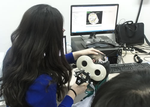
NIRS technology is uniquely positioned to help identify areas of the brain involved in a range of functions - an area of study called "brain mapping." Brain mapping has important applications in and far-reaching implications for a number of fields, from basic science research to diagnosis and treatment of schizophrenia and other psychiatric disorders.
Computerized tomography (CT), functional magnetic resonance imaging (fMRI), positron emission tomography (PET), electroencephalography (EEG) and magnetoencephalography (MEG) are among the most prominent of the technologies used for brain imaging studies. However, these modalities typically suffer from relatively low temporal resolution (CT, fMRI, PET) or spatial resolution (EEG, MEG). Computed tomography and positron emission tomography have the added drawback of deploying ionizing radiation.
NIRS can transcend these limitations, combining excellent temporal resolution (~10 ms) with reasonable spatial resolution. Furthermore, NIRS instruments use safe, non-ionizing radiation and can be made relatively insensitive to head or body motion - an issue not so easily addressed with the technologies noted above.
The technique has already helped to advance basic science brain mapping studies, identifying areas of the brain associated with a range of motor and visual tasks, for example. It might also contribute to the diagnosis and treatment of depression, schizophrenia and Alzheimer's disease. Indeed, researchers are already using NIRS to explore the pathogenesis of these and other psychiatric disorders - taking advantage of its ability to probe the specificity of frontal and prefrontal cortical areas in performing neuropsychological tests in psychiatric patients, for instance.
Visit the Publications page to learn more about studies using NIRS for brain mapping applications.
Read more about our CW6 technology here.


